November 6, 2026
How Standing and Sitting X-Rays Guide Hip Replacement Planning
Standing and sitting X-rays can help with hip replacement planning by showing how your pelvis changes position as you move. That matters because the replacement socket moves with your pelvis, changing its functional position during everyday activities.
A standard hip X-ray shows important details about bone and arthritis. For some patients, comparing positions adds information that helps the surgeon plan for stability. Understanding how the hip and spine work together makes that difference easier to see.
Why Pelvic Movement Matters for a New Hip
Your hip joint connects the thighbone to the pelvis. Above it, the lower spine and pelvis work together to help you stand upright, sit down, and bend. Their movement affects how much motion the hip needs for each activity.
What Usually Happens When You Sit
As many people sit, the pelvis tilts backward and the lower back becomes less arched. That pelvic movement changes the direction the hip socket faces and helps accommodate the bend at the hip.
The amount of movement varies. Hip arthritis, spinal stiffness, and the position you use for the X-ray can all influence what the images show. Your surgeon considers those details rather than treating every patient's movement as identical.
Why the Replacement Socket Moves Too
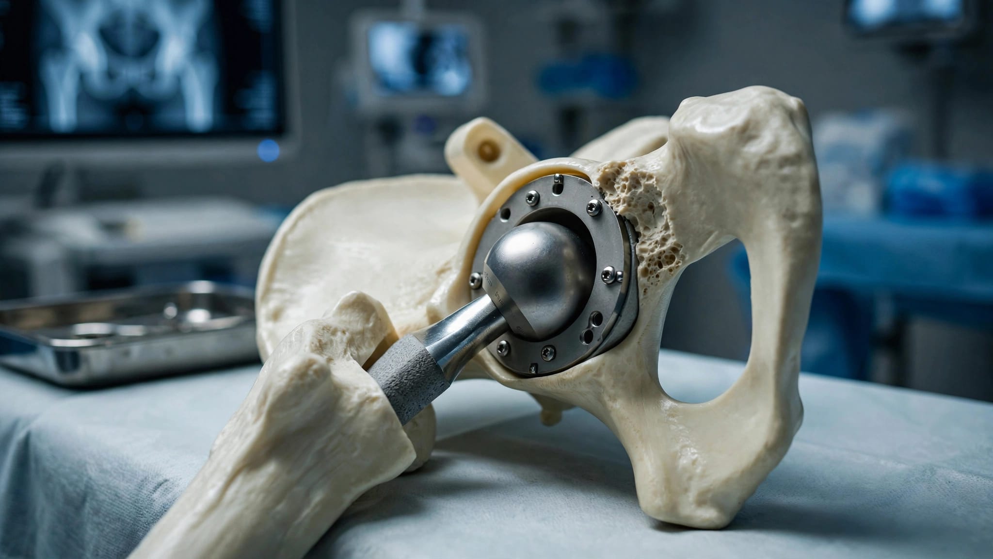
During hip replacement, the surgeon places a cup in the pelvic socket and a stem in the thighbone. A ball on the stem moves inside the cup.
Once implanted, the cup stays fixed to the pelvis. As the pelvis tilts, the cup's orientation relative to the rest of your body changes too. Comparing standing and sitting positions helps the surgeon consider how that cup may function beyond a single posture.
What Standing and Sitting X-Rays Show
Hip replacement imaging often includes a front view of the pelvis, called an anteroposterior or AP view, and a side view of the affected hip. A surgeon may also request side-view X-rays that include the pelvis and lower spine in standing and sitting positions.
The Standing View
A standing image shows your alignment while you bear weight. Depending on the view, it can help the surgeon assess the hip joint, pelvic position, lower spine, and the relationship between both hips.
X-rays can also show joint-space narrowing, bone spurs, deformity, and some differences in leg length. These findings contribute to the plan for fitting and positioning the replacement components.
The Sitting View
A sitting side view gives the surgeon another look at pelvic position and spinal alignment. Comparing it with the standing image shows how much the pelvis changed between the two recorded positions.
Together, the images offer more information about functional pelvic position than either view alone. They are still snapshots, though. They don't show every movement you make while walking, bending, or getting up from a chair.
How Surgeons Interpret the Change Between Positions
The comparison involves more than deciding whether a pelvis moves. The surgeon looks at its position in each image, how much it changes, and how the lower spine and hip contribute to the posture.
Limited Pelvic Movement
If the pelvis moves very little between standing and sitting, the hip may need to bend more to reach the same seated position. After replacement, that extra demand can bring the implant components, or the bones around them, into contact sooner.
That contact is called impingement. In some positions, it can push the ball toward the edge of the cup and increase the chance of instability.
A Pelvis That Moves More
A pelvis that tilts considerably can also change the cup's functional orientation between positions. A cup that appears appropriately positioned on one image may face a different direction when the person stands or sits.
Neither a small nor a large change is a diagnosis by itself. The surgeon interprets the images alongside hip motion, spinal alignment, anatomy, and the planned implant position.
When Spine History Deserves Extra Attention
Standing and sitting views can be useful when a patient's spine or pelvis may not move in the usual way. They can help the surgeon understand the relationship between the two before deciding how to place the cup.
Lumbar Fusion and Spinal Stiffness
A lumbar fusion joins bones in the lower spine and can limit movement in the fused area. Other spinal conditions can also affect flexibility or alignment. However, the effect on pelvic motion differs from person to person.
If you've had spine surgery, your surgeon considers the location of the fusion and your current posture. Our discussion of hip replacement after lumbar fusion explains why that history can affect the cup's functional position.
Hip Stiffness Can Affect the Picture
The hip itself also influences how you sit and stand. Arthritis can restrict hip motion, and pain may change the position you use during an X-ray. A physical exam helps the surgeon understand those limits.
Scoliosis or a change in the curve of the lower back may also affect alignment. The images are most useful when interpreted with your symptoms, exam, and surgical history.
How the Images Influence Component Positioning
A hip replacement plan must account for both secure fixation and movement. The surgeon chooses a position that fits the available bone while considering how the cup will face during daily activities.
Cup Orientation in Daily Positions
The cup has an angle relative to the pelvis, but its functional orientation changes as the pelvis tilts. When the pelvis tilts backward, the cup generally faces more forward. When the pelvis tilts forward, the cup generally faces less forward.
For someone with limited pelvic movement, the surgeon may adjust the planned orientation to account for that pattern. The choice also depends on the shape of the socket and where the cup can obtain reliable support. One fixed target doesn't fit every hip.
The Cup and Stem Work Together
The cup is only one part of the reconstruction. The rotation and position of the femoral stem also affect how the ball moves inside the socket. Leg length, hip offset, and soft-tissue tension matter for stability and muscle function.
Surgeons use preoperative templating for hip replacement to estimate component sizes and positions. Those estimates guide the operation, but the final choices can change when the surgeon assesses the bone and tests the hip during surgery.
What the Findings Mean for Dislocation Risk
A dislocation occurs when the replacement ball comes out of the socket. Cup position and pelvic movement are part of that risk, along with muscle support, implant design, surgical history, and other factors.
Why Movement Can Affect Stability
When a person with limited backward pelvic tilt sits down, the cup may not gain as much forward-facing orientation as expected. The hip may need more flexion, which can increase the chance of impingement and instability in that position.
Other alignment patterns can create different concerns when standing or moving into extension. Standing and sitting X-rays help identify these patterns, but they can't predict with certainty whether a dislocation will occur.
When a Different Implant May Be Considered
For some patients with higher instability risk, a surgeon may discuss a dual mobility implant. This design uses a small ball within a larger mobile liner and can provide more movement before impingement.
A spine condition doesn't automatically mean you need this option. Our guide to dual mobility hip implants covers who may benefit and the implant-specific risks. No implant design eliminates the possibility of dislocation.
What to Expect During the X-Rays
For a standing view, a radiologic technologist helps position you beside the imaging equipment. For a sitting view, you'll sit as directed while the image is taken. The requested posture may vary with the surgeon's planning needs.
Tell the Team About Movement Limits
If standing is difficult or sitting causes pain, tell the imaging team. They can help you understand the requested position and work with you within your limits. A painful or unusual posture can affect how the image represents your everyday alignment.
You'll need to remain still briefly for each exposure. X-rays use a small amount of ionizing radiation, so tell the team if you're pregnant or may be pregnant before the study.
Why You May Need Other Images
Standing and sitting views don't replace every other test. A CT scan may help show complex bone anatomy, while other imaging may be useful for certain soft-tissue concerns.
For example, planning around existing hip hardware may require additional views of the femur or a closer look at old plates and screws. The imaging choice depends on the question your surgeon needs answered.
Discussing Your Imaging With a Hip Surgeon
You don't need a detailed knowledge of X-ray measurements to have a useful consultation. Ask your surgeon what the images show about your pelvic movement and whether that finding changes the proposed plan.
Mention previous spine or hip operations, prior dislocations, and difficulty sitting or standing. If you have records from an earlier operation, bring them. They can help your surgeon understand existing implants or a spinal fusion.
It's also reasonable to ask how the planned cup position and implant choice address your movement pattern. At our Fort Myers practice, we provide hip replacement care and can discuss how your anatomy and imaging fit into your treatment options.
A Clearer Picture of Your Hip in Motion
Standing and sitting X-rays give a surgeon two views of how your pelvis and spine relate to your hip. That comparison can add useful context when planning component position and considering stability.
The images are one part of a personalized plan . Your exam, medical history, bone anatomy, and goals matter alongside what appears on the X-rays.
ADDITIONAL ARTICLES