November 15, 2026
Femoral Version in Hip Replacement Planning
Femoral version is the rotational alignment of the upper thighbone, and it helps guide how a surgeon positions a hip replacement. It matters because the new ball and socket must work together as you sit, stand, walk, and turn.
Your anatomy may allow a straightforward reconstruction, or it may call for adjustments to the implant plan. Understanding rotation can help you have a more informed conversation with your surgeon.
What Femoral Version Means
The femur is your thighbone. At its upper end, the femoral neck connects the shaft to the ball of the hip. Femoral version describes how that neck points in relation to the lower end of the femur, near the knee.
Surgeons describe version as an angle, but you don't need to know your measurement to understand the basic idea. The direction of the femoral neck affects how the hip meets the socket and moves through everyday positions.
Anteversion and Retroversion
With femoral anteversion, the femoral neck points forward relative to the lower femur. People naturally have different amounts of forward rotation.
Retroversion describes a neck that points backward relative to that reference. Some hips also have less forward rotation than expected without being truly retroverted. Both patterns are part of the anatomy a surgeon considers.
Natural Bone and Implant Rotation
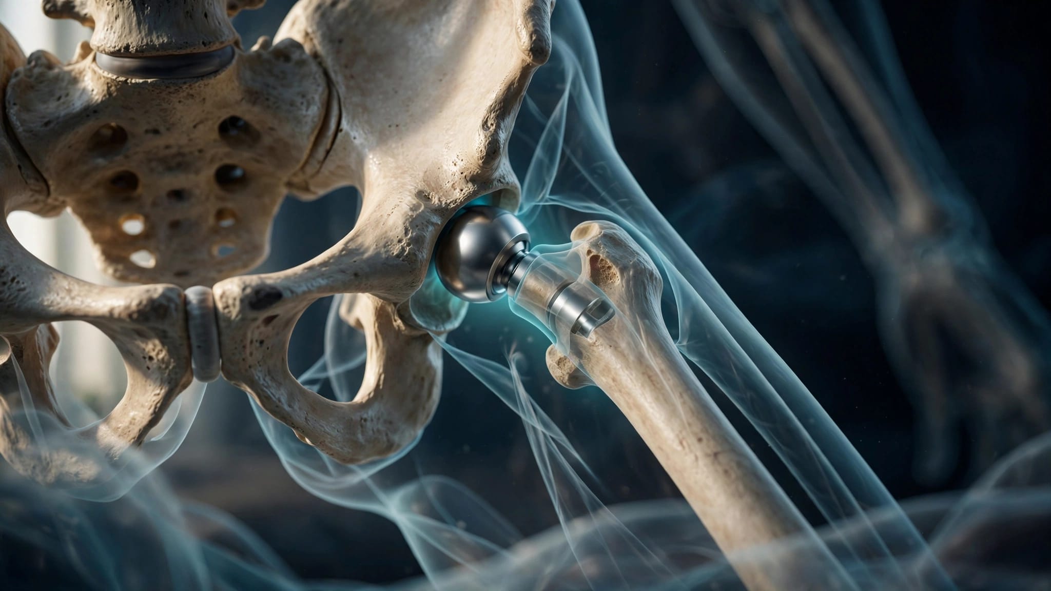
During a total hip replacement, the surgeon removes the damaged femoral head and places a stem inside the thighbone. A new ball attaches to that stem.
The femoral component's version describes the orientation of the implanted neck and ball relative to the lower femur. It may differ from the version of your natural bone. The aim is to achieve stable fixation and a functional relationship with the new socket, rather than reproduce one measurement in isolation.
How Rotation Affects Movement and Stability
Your replacement hip needs enough room to move without its parts contacting each other prematurely. It also needs support from the surrounding muscles and soft tissues.
Impingement and Range of Motion
Impingement occurs when structures make contact and limit movement. After hip replacement, the implant neck can contact the edge of the socket liner. Bone can also contact bone during certain movements.
Early contact may make actions such as bending to sit or turning the leg more difficult. In some positions, it can also create a levering force on the joint. Component orientation, implant design, bone shape, and soft-tissue tension all influence how much motion is available.
Dislocation Risk
A dislocation occurs when the replacement ball comes out of the socket. The relationship between the cup and stem can influence stability, especially near the limits of movement.
A hip with excessive combined forward orientation may be more vulnerable to instability in extension and outward rotation. Too little forward orientation may increase the chance of instability during flexion and inward rotation. These are mechanical considerations, not predictions of what will happen to a particular patient.
Muscle strength, prior surgery, falls, and healing also matter. Careful alignment can reduce avoidable mechanical problems, but it cannot eliminate every risk.
How Surgeons Assess Femoral Version
Planning begins with your symptoms, medical history, and a physical examination. Imaging adds information about bone shape and helps the surgeon prepare for implant placement.
Physical Examination and X-Rays
During the exam, your surgeon may assess how far your hip rotates inward and outward. Walking pattern, muscle strength, leg length, and stiffness also help build a picture of how your hip functions.
Rotation on examination doesn't provide an exact measurement of femoral version. Pain, arthritis, and tight tissues can all affect the movement your surgeon observes.
Standard imaging often includes a pelvis X-ray and another view of the affected hip. X-rays show arthritis, overall bone shape, leg-length differences, and previous hardware. They also support templating , a process that estimates implant sizes and positions before surgery. However, ordinary X-rays don't always show rotational anatomy clearly enough to measure femoral version.
When CT Adds Useful Detail
A CT scan can provide a more detailed view of the femur and socket. To measure femoral version, the imaging assessment can compare the orientation of the femoral neck with a reference near the knee.
CT may be helpful when anatomy is difficult to interpret, particularly after a fracture or previous surgery. It can also help assess bone loss and hardware. It isn't necessary for every hip replacement, so the surgeon weighs its potential value against factors such as radiation exposure.
How Femoral Version Works With Cup Orientation
The acetabulum is the socket on the pelvic side of your hip. During replacement, the surgeon positions an artificial cup in that socket. The cup's orientation matters alongside the direction of the femoral stem.
Understanding Combined Anteversion
Cup anteversion describes how much the socket opening faces forward. Combined anteversion is a way of considering the forward orientation of both the cup and femoral component together.
The concept helps the surgeon assess how the two parts will meet during movement. However, adding two measurements doesn't capture every detail of a three-dimensional joint. Cup tilt, pelvic position, implant design, and soft tissues also affect how the hip functions.
There isn't one combined angle that fits every patient. The surgeon must also consider whether the cup has adequate bone support and whether the stem can be fixed securely.
Why Both Components Need a Plan
A stem that fits the femur well may settle in an orientation influenced by the shape of the canal. The surgeon may then need to account for that orientation when positioning the cup.
Likewise, socket bone loss or an unusual socket shape may limit where the cup can safely sit. In that situation, the femoral side of the reconstruction becomes part of the discussion. Our guide to hip implant sizing and component alignment explains how positioning works alongside implant size, leg length, and offset.
Why Pelvic and Spine Movement Matter
Your pelvis doesn't stay in exactly the same position throughout the day. As you sit, it usually tilts backward, changing how the socket faces relative to your femur. Standing brings the hip into a different position.
A stiff spine or previous spinal fusion can limit that adjustment. When the pelvis moves less, the hip may need to accommodate more movement during activities such as sitting down. This can change the mechanical demands on a replacement joint.
For patients with significant spinal stiffness or a history of fusion, the surgeon may consider standing and sitting images as part of planning. The goal is to understand how the pelvis behaves during daily activities, rather than rely only on its position in a single image.
A spine condition doesn't automatically mean a patient needs a different implant. It does give the surgeon another factor to consider when assessing component orientation and stability.
When Hip Anatomy Calls for Extra Planning
Many people have some variation in femoral version. More pronounced changes, bone loss, or previous procedures can make implant placement less predictable.
Hip Dysplasia
Hip dysplasia can involve a shallow socket, altered femoral rotation, or a narrower femoral canal. The surgeon must consider both sides of the joint: the cup needs support from pelvic bone, and the stem needs a secure fit in the femur.
The plan may also address leg length and hip offset, the distance that helps position the femur relative to the center of the joint. These features affect muscle tension and walking mechanics.
Dysplasia doesn't automatically rule out a particular surgical approach. Our discussion of hip replacement planning for dysplasia covers how socket and femoral anatomy can affect that decision.
Fractures and Previous Surgery
A healed fracture can change the shape or rotation of the femur. On the pelvic side, a previous socket fracture may leave irregular bone or hardware near the planned cup. The surgeon considers those changes when deciding where components can gain stable support.
Previous rods, plates, or screws may also affect access to the femoral canal. Some hardware can remain in place, while other pieces may need removal. Our guide to hip replacement around existing hardware describes how that history can change the operation.
How Rotation Influences Surgical Decisions
A preoperative plan is a starting point. During surgery, the surgeon assesses the available bone, prepares the joint, and tests how the proposed components fit together.
Stem Fit and Component Position
Different femoral stem designs fit and engage the bone in different ways. Some may offer more options for adjusting rotational position, while others are guided more closely by the shape of the femoral canal.
A surgeon won't sacrifice secure fixation just to force a stem toward a preferred angle. Instead, the plan may involve a different implant or an adjustment elsewhere in the reconstruction. Cup position still needs sufficient bone coverage and a stable fit.
Trial components allow the surgeon to assess movement, soft-tissue tension, leg length, and stability before the final components are secured. Findings during surgery may lead to a change in the original plan.
Implant Design and Surgical Approach
For some patients at higher risk of instability, a surgeon may discuss a dual mobility implant. This design includes an additional bearing surface that can provide more movement before impingement. It may improve stability for selected patients, but it doesn't replace careful component positioning or remove all risk.
The surgical approach is a separate decision. SuperPATH is a route used to access the hip; it doesn't determine the implant's version by itself. Your surgeon needs enough access to prepare the bone and position the components accurately.
Technology can assist with planning or placement in some cases. Our comparison of SuperPATH and robotic hip surgery explains the distinction between an approach and a planning tool.
What to Discuss With Your Surgeon
When you're considering hip replacement, ask how your anatomy affects the proposed operation. If you have dysplasia, a previous fracture, spinal fusion, or prior hip surgery, tell your surgeon about it. Older images and operative reports can be useful when available.
You can also ask whether your femoral or socket anatomy changes the choice of implant, whether additional imaging would help, and how the surgeon assesses stability during surgery. The answers should connect the plan to your needs, rather than promise a particular outcome.
After surgery, follow the movement and recovery guidance your team gives you. If you develop sudden severe hip pain, an apparent deformity, or can't use the leg after a fall or twist, seek urgent medical care. These can be signs of a dislocation or fracture.
A Plan Built Around Your Hip
Femoral version is one part of a larger hip replacement plan. Its relationship with the socket, your bone shape, and your everyday movement helps guide component placement.
A careful assessment gives your surgeon a way to balance stable fixation with comfortable movement. If you're considering hip replacement, discuss how your own anatomy shapes the options available to you.
ADDITIONAL ARTICLES