August 26, 2026
MRI After Hip Replacement: Implant Safety and Scan Quality
An MRI appointment can feel routine until a screening form asks about your hip replacement. If you need an MRI after hip replacement , don't treat that question as routine paperwork. The exact implant, its labeling, and the planned scanner settings can affect whether the exam proceeds.
MRI is often possible for people with modern hip implants, but "hip replacement" alone cannot clear a scan. Metal can also blur the area under review, so an exam that meets safety conditions may still offer limited answers. Your treating clinician and radiology team decide which study fits your symptoms and surgical history.
Good records and a thoughtful protocol help them make that decision with fewer surprises.
MRI After Hip Replacement Starts With Implant Details
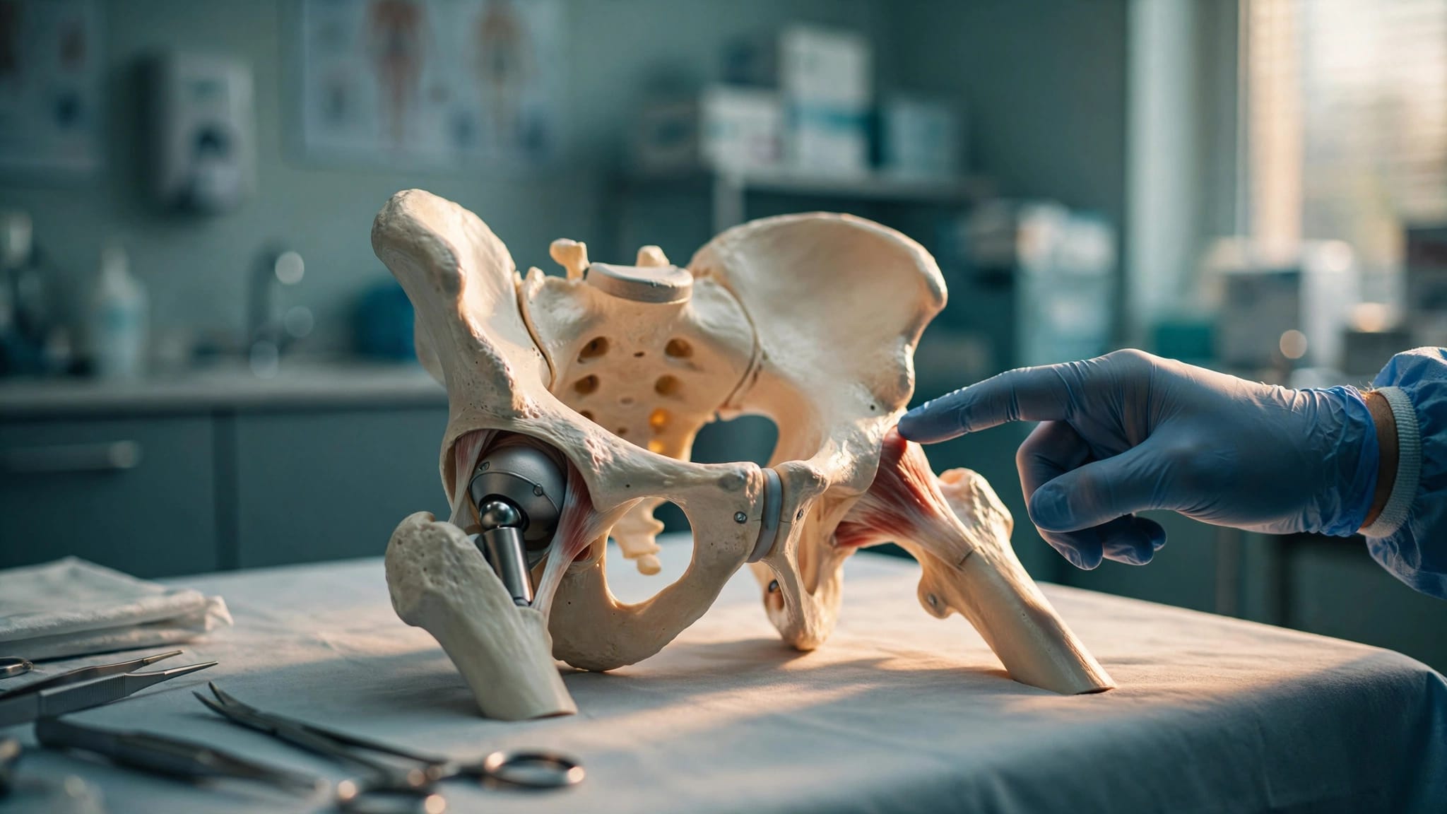
A hip replacement is not one standard device. Brand, component design, materials, and the details of earlier surgery all matter. Your implant could include a titanium stem, cobalt-chromium parts, a ceramic head, a polyethylene liner, screws, or several of these materials.
The radiology team needs device details, not an estimate based on your surgery date or hospital. If your operation was recent, your surgeon may also consider incision healing, comfort while lying flat, and whether imaging will change treatment.
A total hip has several components
Most total hip systems are passive implants, which means they do not contain a battery or programmed electronics. That distinction matters, although it does not give automatic MRI clearance.
Revision components, retained fracture hardware, older surgery, and uncommon devices may change the review. A replacement hip also differs from a pump, neurostimulator, or pacemaker, which can carry separate MRI restrictions.
Your orthopedic office may have the operative report or implant sticker sheet in your chart. If another surgeon performed the procedure, the hospital records department may be able to provide those details.
MR Conditional means conditions apply
MRI device labels commonly use MR Safe, MR Conditional, and MR Unsafe. An MR Safe device presents no known hazard in the MR environment. An MR Conditional device can be scanned only under listed conditions, while an MR Unsafe device must stay out of the MR environment.
Those instructions may cover magnetic field strength, radiofrequency energy limits, gradient settings, body position, and scan length. A 1.5 T scanner and a 3 T scanner do not always produce the same conditions. This is why trained MRI staff must clear the exam rather than relying on a general online answer.
Implant Records Help the Team Clear the Scan
The American College of Radiology calls for positive written or electronic identification of implanted devices before MRI. That process protects you from a decision based on an incomplete memory of what was placed during surgery.
A previous MRI without a problem is useful history. Still, it does not replace confirming the implant and the conditions for the new scan.
Documents fill in missing details
Bring your implant card if you have one. It may list the manufacturer, product name, catalog number, model number, and implantation date. An operative report, implant sticker sheet, patient portal record, or hospital medical-records contact can also help.
Tell the ordering clinician where and when you had hip replacement surgery if you no longer have records. The imaging team may then review manufacturer labeling or request documentation before scheduling the study.
A photo of your implant card can be helpful. However, staff may still need the complete report when the card does not identify every component.
Screening considers your whole medical history
Before your exam, answer every screening question carefully. Mention other surgeries, retained bullets or shrapnel, aneurysm clips, cochlear implants, implanted pumps, stimulators, or any metal injury to an eye.
Also tell the team about kidney disease, prior contrast reactions, pregnancy, claustrophobia, and trouble lying flat. These concerns do not always rule out MRI, but they may alter the plan.
A cleared hip implant does not clear every other implant in your body. Do not bring a phone, keys, hearing aids, credit cards, or removable metal into the scan area until MRI staff has checked them.
Implant Safety and Image Quality Are Different Issues
The words "safe for MRI" and "clear MRI picture" describe separate questions. Implant safety concerns how the device behaves in the MR environment. Image quality concerns whether the radiologist can see the tissue or bone related to your symptoms.
Both questions matter, especially when new pain develops after a hip replacement.
What the MRI team assesses for safety
Metal can interact with magnetic and radiofrequency fields. The team reviews possible force or twisting, radiofrequency heating, and small electrical currents that conductive materials can develop.
With many modern passive orthopedic implants, clinically meaningful movement is uncommon. Yet an uncommon risk is still a reason to follow the implant's stated conditions and the facility's screening process.
During the scan, stay in contact with the technologist. Report unexpected heating, burning, pain, or discomfort right away. Cables, blankets, and skin position also need attention because direct contact points and closed skin loops can contribute to burns.
Metal artifact is often the practical limit
Artifact is usually the bigger problem near a prosthetic hip. Metal disrupts the MRI signal and can create warped anatomy, dark areas, bright signal pileup, and missing detail near the joint.
That distortion may hide a fluid collection, tendon injury, bone change, or tissue reaction. It can also extend into images of the pelvis and upper thigh.
Artifact depends on the implant's material and size, its orientation, the magnet strength, and the imaging sequence. An MRI after hip replacement may be permitted under the implant's conditions yet still be a poor test for the clinical question.
MRI Protocols Can Reduce Metal Artifact
A detailed imaging order helps the radiology team build a more useful protocol. The ordering clinician should explain the concern, such as new groin pain, a suspected abductor tendon injury, a possible fluid collection, or a soft-tissue problem near the joint.
That information guides the choice of sequences and the body area included in the exam.
Routine settings can make a meaningful difference
Many centers use MARS MRI, a broad term for metal artifact reduction methods. The protocol often favors fast spin-echo or turbo spin-echo imaging because gradient-echo sequences tend to create more distortion near metal.
Technologists can also adjust echo time, receiver bandwidth, slice thickness, and image orientation. A lower field strength, often 1.5 T rather than 3 T, may reduce susceptibility artifact around a hip prosthesis.
However, lower field strength is not automatically better for every exam. The radiologist weighs artifact reduction against the detail needed for the body part under review.
SEMAC and MAVRIC can improve local views
Some scanners offer dedicated sequences called SEMAC, short for slice encoding for metal artifact correction, and MAVRIC, short for multi-acquisition variable-resonance image combination. Both target distortion that standard sequences cannot fully correct.
These methods may improve views of tissues near the cup, stem, and surrounding muscles. They can also make the scan take longer, and results still vary with the implant, scanner, and reason for imaging.
If your surgeon orders MRI for a complex concern near a replacement hip, ask whether an imaging center experienced with orthopedic metal-reduction MRI is appropriate. The right protocol can make the difference between a vague result and an actionable one.
When X-Ray, CT, or Ultrasound May Fit Better
MRI is useful when soft tissues need close evaluation. Still, it is not automatically the first test after hip replacement. The right exam depends on whether the concern involves alignment, fracture, loosening, bone loss, infection, fluid, muscles, or a problem farther from the implant.
Your symptoms and examination guide that choice.
X-rays and CT provide strong bone detail
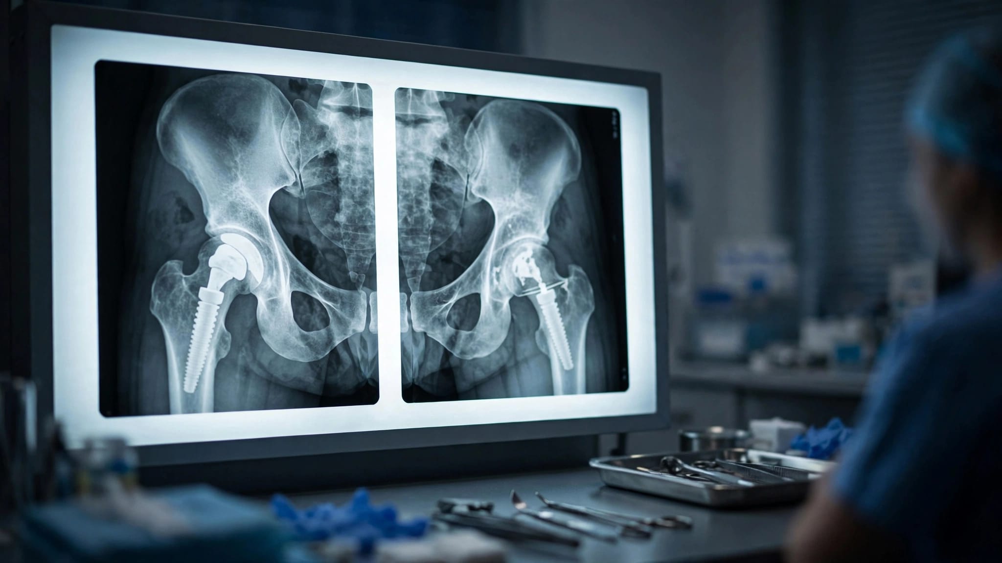
Standard X-rays often begin the evaluation. They can show component position, dislocation, visible fracture, alignment, and some signs of loosening or wear.
For bone detail, component version, osteolysis, or fracture questions that X-rays cannot settle, CT may help. Modern CT also offers metal artifact reduction methods, although metal can still limit parts of the image.
Patients with retained plates or screws after past trauma may need CT imaging for hip hardware assessment when the surgeon needs a clearer view of bone and existing hardware.
Ultrasound can assess fluid and selected soft tissues
Ultrasound does not create metal artifact. It can help evaluate a superficial fluid collection, trochanteric bursitis, or selected tendon concerns. It can also guide an aspiration when infection is suspected.
Your clinician may combine ultrasound findings with blood tests, X-rays, and fluid analysis rather than rely on one test alone. Nuclear medicine studies may also be considered in selected cases, though results need careful interpretation after joint replacement.
MRI has a different strength. When the implant is cleared and artifact reduction can answer the question, it may show muscles, tendons, nerves, and deep soft tissue that X-ray and CT cannot show as well.
A Clear Plan Leads to Better Imaging Decisions
A hip replacement does not automatically prevent MRI, but it does require careful screening. The implant's exact identity and its scan conditions matter more than assumptions about the surgery.
Bring any implant records you can find, describe all other medical devices, and share your symptoms clearly. That gives your orthopedic clinician and radiology team the information needed to choose a safe, useful study.
The best test is the one that answers the clinical question while respecting your individual implant and medical history .
ADDITIONAL ARTICLES