September 6, 2026
Hip Arthritis MRI: When an X-Ray Isn't Enough
Hip pain can make every step feel negotiated, especially when stiffness affects sleep, work, or simple tasks like putting on shoes. A hip arthritis MRI can reveal problems an X-ray cannot see, but it isn't the starting test for every painful hip.
For many people, plain X-rays provide the information needed to confirm osteoarthritis and begin treatment. MRI becomes more useful when symptoms, examination findings, and X-rays don't tell the same story.
Why Hip X-Rays Usually Come First
X-rays are fast, widely available, and excellent at showing the bones that form the hip joint. Most orthopedic evaluations begin with an X-ray of the pelvis and one or more views of the painful hip.
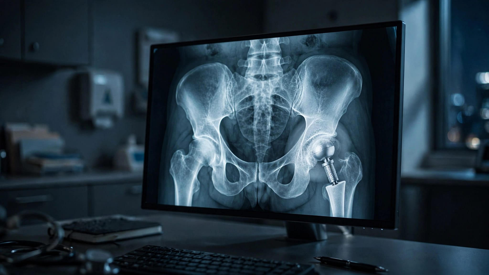
The images can show joint-space narrowing, bone spurs, cysts in the bone, changes in joint shape, and severe "bone-on-bone" wear. They can also reveal an obvious fracture, a dislocation, or arthritis caused by an old injury.
X-rays show cartilage loss indirectly
Cartilage does not appear on a standard X-ray. Instead, the space between the femoral head and the socket gives an indirect clue about how much cartilage remains.
When osteoarthritis advances, that gap often becomes narrower. Still, the degree of narrowing does not measure pain perfectly. Someone may have marked arthritis on film yet remain fairly active, while another person with modest changes may have major limits.
For a closer look at phrases that often appear in reports, review these hip arthritis X-ray results.
Many treatment decisions don't require MRI
When the history, physical examination, and X-ray all point to hip osteoarthritis, an MRI may not change the next step. Treatment can include activity changes, physical therapy, medication guidance, injections, or a discussion of joint replacement when pain and function warrant it.
An MRI is not a test to order simply because an X-ray shows arthritis. The scan should answer a focused clinical question that could change care.
When a Hip Arthritis MRI Adds Detail
A hip arthritis MRI looks beyond the bone outlines. It shows cartilage, the labrum, tendons, muscles, fluid, and bone marrow. That broader view helps when an orthopedic surgeon suspects another source of pain or an additional injury.
A hip arthritis MRI is often considered when pain seems stronger than expected from the X-ray, symptoms began suddenly, or the physical exam points to a soft-tissue or bone marrow problem.
Cartilage and labral damage
The labrum is a rim of fibrocartilage around the hip socket. It helps deepen the socket and contributes to joint stability. A labral tear can cause groin pain, clicking, catching, or pain during twisting, although some tears cause no symptoms at all.
MRI can also show focal cartilage injury before joint-space narrowing becomes obvious on X-ray. In selected cases, a clinician may order an MR arthrogram, which involves injecting contrast into the joint before imaging, when a labral tear is a major concern.
These findings matter most when they match your symptoms and examination. A scan alone cannot prove that a labral tear is causing pain.
Tendons and muscles around the hip
Outer hip pain may come from gluteal tendons, nearby bursae, or muscle injury rather than arthritis inside the joint. MRI can identify tendon tearing, tendinopathy, muscle swelling, and fluid around the greater trochanter.
Pain near the buttock may also arise from the lower back or sacroiliac joint. Location can mislead because pain often travels. A careful comparison of hip arthritis versus back pain can help explain why the examination matters before selecting a scan.
Bone Marrow Changes Can Explain Severe Pain
MRI detects changes inside bone that plain films may miss. Bone marrow edema is a descriptive MRI finding that can appear after stress injury, trauma, arthritis flare-ups, inflammatory conditions, or other problems.
It is not a diagnosis by itself. Your surgeon or radiologist must interpret marrow changes alongside symptoms, medical history, and other images.
Occult and stress fractures
An occult fracture is a break that does not appear clearly on an initial X-ray. This concern is especially important after a fall, even if the person can still take a few steps.
MRI can find fractures of the femoral neck, pelvis, or nearby bone before they become visible on X-ray. It can also identify stress injuries caused by repetitive loading. Runners can develop these injuries, but so can people with osteoporosis, sudden increases in walking, or weakened bone.
A possible fracture needs timely evaluation because continuing to bear weight may worsen some injuries. CT can also help answer certain fracture questions, especially when a surgeon needs fine bone detail.
Avascular necrosis of the femoral head
Avascular necrosis, also called osteonecrosis, occurs when part of the femoral head loses its blood supply. Early in the process, an X-ray may look normal or show little change.
MRI is highly useful for detecting suspected avascular necrosis before the rounded femoral head collapses. Risk factors can include prior high-dose corticosteroid use, heavy alcohol exposure, some blood disorders, prior hip trauma, and certain medical treatments. However, the condition can also occur without an obvious cause.
Early identification may affect treatment options, which is why persistent groin pain with an unrevealing X-ray sometimes calls for further imaging.
MRI Helps Separate Osteoarthritis From Other Hip Problems
Osteoarthritis commonly causes deep groin pain, stiffness after rest, reduced hip rotation, and pain with walking or standing. Yet hip pain has many possible causes, and arthritis visible on an X-ray may be only part of the picture.
For example, bursitis tends to produce tenderness on the outer hip. Lumbar nerve problems may bring pain below the knee, numbness, tingling, or weakness. Inflammatory arthritis, tendon injury, hernia, pelvic conditions, and referred pain can also resemble hip-joint pain.
Infection needs prompt attention
MRI can show fluid, inflammation in soft tissues, and bone changes that may raise concern for infection. Still, MRI does not diagnose a joint infection on its own.
If infection is suspected, an orthopedic clinician may combine the scan with blood tests and a joint aspiration. Ultrasound can guide aspiration when fluid is accessible. The lab analysis of that fluid often provides information imaging cannot.
Seek prompt medical attention for a hot or red hip, fever, chills, rapidly worsening pain, or an inability to bear weight. These symptoms need assessment rather than waiting for a routine imaging appointment.
Symptoms that should not wait
Contact a medical professional promptly after a fall if hip pain makes walking difficult, even if an early X-ray seems reassuring. New weakness, loss of bladder or bowel control, or numbness in the groin also needs urgent evaluation.
Sudden shortness of breath, chest pain, fainting, or coughing blood require emergency care. Those symptoms can point to conditions outside the hip but should never be ignored.
MRI for Surgical Planning Has a Defined Role
For straightforward hip replacement caused by advanced osteoarthritis, X-rays usually provide the main planning images. They show joint wear, leg-length differences, bone shape, alignment, and overall bone quality.
MRI may add useful detail if there is concern about abductor tendon damage, a tumor-like bone lesion, infection, avascular necrosis, or unusual soft-tissue anatomy. It may also help when a painful hip has symptoms that don't fit the degree of arthritis shown on X-ray.
Prior trauma or hardware may need another scan
People who have had pelvic fractures, hip surgery, plates, screws, or other retained hardware may need additional imaging before replacement. In these cases, CT often gives a clearer map of bone loss, healed deformity, and the relationship between hardware and the planned implant.
MRI can be difficult near metal because the hardware distorts the image. Some facilities use metal-artifact reduction techniques, often called MARS MRI, to improve views of nearby muscle, tendons, fluid collections, and other soft tissues.
Complex cases call for an individualized plan. Information about hip replacement after pelvic fracture explains why prior trauma can change the imaging and surgical discussion.
Why MRI Is Not Routine for Every Painful Hip
MRI takes longer than X-ray, costs more, and requires you to lie still in a narrow scanner. Claustrophobia, severe pain while lying flat, and certain implanted devices can make the test harder, although many modern implants are MRI-conditional after proper screening.
Contrast is not necessary for every hip MRI. When contrast is considered, the ordering clinician reviews kidney function, prior reactions, and the question the scan needs to answer.
Incidental findings can complicate the picture
MRI is sensitive, which can be helpful and frustrating. It may reveal a small labral tear, tendon wear, cyst, or marrow change that has nothing to do with your symptoms.
Age-related changes are common, even in people without hip pain. Treating every abnormal phrase on an MRI report can lead to unnecessary worry or procedures. The useful question is whether a finding matches the pain pattern, exam, and daily limitations.
A hip arthritis MRI works best as part of a full orthopedic evaluation, not as a substitute for one.
The Right Scan Starts With the Right Question
If your hip pain is persistent, worsening, or changing the way you walk, bring prior imaging and a clear symptom history to your appointment. Note where the pain starts, what movements trigger it, whether it wakes you at night, and whether a fall or injury came before it.
X-rays often answer the central question in osteoarthritis. MRI earns its place when there may be hidden fracture, early osteonecrosis, infection, cartilage or labral injury, or a soft-tissue problem that could change treatment.
The most useful image is the one that helps explain your symptoms and supports a sound plan for getting you moving more comfortably.
ADDITIONAL ARTICLES