August 22, 2026
SuperPATH pelvic fracture: Planning After Hip Trauma
A healed pelvic fracture can continue to shape hip replacement years later. If you are considering a SuperPATH pelvic fracture case, the main question is whether the surgeon can restore the hip safely with the available bone, anatomy, and surgical exposure.
Prior acetabular fixation, altered hip alignment, scar tissue, bone loss, and poor bone quality can change the imaging, implant choice, and surgical plan. Some patients remain good candidates for SuperPATH. Others need a different approach that provides more control. The decision starts with the details of your hip, not the fracture history alone.
Why a prior pelvic fracture changes hip replacement planning
A pelvic fracture and an acetabular fracture are related but not identical. A pelvic injury may involve the pubic rami, ilium, sacrum, or pelvic ring. An acetabular fracture affects the socket that holds the femoral head.
The injury's location, severity, alignment, and treatment all influence a later hip replacement. Even after healing, the hip may have post-traumatic arthritis, a changed hip center, uneven socket walls, or a leg-length difference. Screws or plates may also sit near the planned acetabular cup.
The original injury matters
The surgeon will want more than the phrase "pelvic fracture" in your medical history. Operative reports, old CT scans, implant records, and information about infection treatment can show which walls or columns were involved.
If those records aren't available, current X-rays and CT imaging can still show much of the present anatomy. However, they may not reveal the original fracture pattern or earlier complications. Tell the surgeon about wound drainage, repeat operations, blood transfusions, and any treatment for infection.
A fracture history doesn't automatically exclude SuperPATH
Some patients with a healed fracture have enough bone and a workable socket shape for a tissue-sparing approach. Others need wider exposure for hardware removal, deformity correction, bone grafting, or stable implant fixation.
SuperPATH hip replacement candidacy depends on the complete picture, including symptoms, motion, strength, imaging, bone stock, medical health, and the technical demands of the operation.
SuperPATH pelvic fracture candidacy starts with anatomy
SuperPATH is designed to limit disruption to muscles and other soft tissues around the hip. That design may support early movement for selected patients, but it also gives the surgeon less direct exposure than some conventional approaches.
In routine arthritis, the surgeon may see predictable landmarks and adequate socket bone. Previous trauma can make those landmarks less reliable. The procedure still requires accurate preparation of the acetabulum and femur, stable component fixation, and control of leg length and hip stability.
The physical exam adds information
The exam includes hip motion, contractures, pain location, leg length, gait, abductor strength, and the condition of the opposite hip. A stiff hip with a fixed deformity may require more access than a hip with mild motion loss.
The surgeon also checks the skin and soft tissues around old incisions. Prior scars don't automatically prevent SuperPATH, but they may affect positioning, exposure, and wound planning.
The reason for replacement affects the plan
Post-traumatic arthritis can produce different bone loss and deformity than primary osteoarthritis. A previous dislocation, infection, nonunion, or malunion adds further planning concerns.
Much of the published SuperPATH research involves routine primary hip replacement rather than patients with previous acetabular fixation. Comparative studies have not shown a consistent pain or functional advantage over standard approaches. A smaller incision should not outweigh the need for reliable reconstruction.
Imaging maps the socket, hardware, and femur
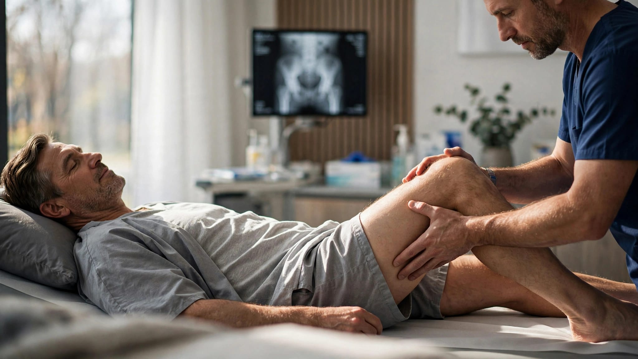
Plain X-rays usually start the workup. An AP pelvis view and hip views can show joint-space loss, the hip center, leg-length differences, hardware position, pelvic alignment, and general bone quality. Comparing both hips and reviewing older films can show how the anatomy changed.
The purpose is not only to confirm arthritis. Imaging helps determine whether the planned cup can sit in viable bone and whether the femoral component can achieve stable fixation.
X-rays support surgical templating
Preoperative templating estimates cup size, stem size, hip center, offset, and the correction needed. With a prior fracture, the surgeon may account for pelvic rotation, healed displacement, metal overlap, and an uneven acetabular rim.
A standard target for cup inclination or version may not fit a damaged socket. The surgeon must balance orientation with the bone that remains available and with the position of the femur.
CT can show what overlapping metal hides
CT may help map retained screws and plates, acetabular version, column integrity, wall defects, and areas of bone loss. It can also show whether a screw enters the region needed for reaming or cup fixation.
CT isn't necessary for every patient with a prior pelvic fracture. There is no universal SuperPATH-specific CT protocol for these cases. The decision depends on the fracture pattern, hardware, X-ray quality, symptoms, and the planned reconstruction. Metal artifact can still limit detail, so CT findings must be reviewed with the radiographs and physical examination.
Retained hardware and bone quality can change implant choices
Old fixation is one of the first issues patients ask about. The answer depends on where the implants sit and whether they interfere with the new hip.
Hardware may stay when it doesn't block reconstruction
Pelvic plates and screws don't always need removal. If an implant sits away from the cup, doesn't prevent reaming, and doesn't interfere with screw placement, leaving it in place may avoid additional dissection.
Removal becomes more likely when hardware blocks the cup, occupies a planned screw path, prevents access to stable bone, or shows signs of loosening or infection. Taking out a well-fixed implant can add scar dissection, blood loss, and fracture risk. Post-traumatic hip replacement literature generally supports removing hardware only when it is necessary for the reconstruction or treatment of a problem.
Bone stock affects fixation
A healed fracture may leave enough bone for a standard press-fit cup and stem. Osteoporosis, disuse, prior infection, bone loss, or a thin acetabular wall can reduce fixation strength.
Poor bone quality raises the risk of a crack or fracture during femoral preparation, stem insertion, acetabular reaming, or cup impaction. The surgeon may change the implant size, fixation method, or planned level of fixation. Options may include additional acetabular screws, another cup design, cemented fixation, or a stem that obtains fixation farther down the femur.
Published SuperPATH reports include femoral fractures, an intraoperative acetabular fracture, and later acetabular fixation failure. These reports don't predict an individual outcome, but they show why bone quality and backup implants belong in the planning discussion.
Component positioning requires more than a standard target
Hip replacement works best when the cup and stem match the patient's anatomy, soft-tissue tension, and movement pattern. Prior fracture can make those goals harder because the original hip center or socket walls may no longer be normal.
The cup must fit the available bone
The surgeon plans cup inclination, anteversion, depth, hip center, and screw direction. A cup placed too high, too vertical, or too far forward or backward can affect stability, leg length, wear, and contact with surrounding bone.
A familiar radiographic safe zone is a useful reference, but it cannot replace three-dimensional assessment. A post-traumatic socket may require a compromise between ideal orientation and secure contact with living bone. Screw direction must also protect surrounding pelvic structures.
The femoral side needs equal attention
SuperPATH studies report acceptable cup positioning in experienced hands, but researchers have assessed cup position more often than stem position. Prior trauma, altered femoral loading, or abnormal version can make femoral preparation less predictable.
The surgeon checks femoral version, canal shape, offset, and leg length during the procedure. If a press-fit stem doesn't feel stable, another fixation strategy may be safer. Intraoperative imaging or other verification methods may help confirm the reconstruction, but they don't replace surgical judgment.
When SuperPATH exposure may be limited
The main issue is whether the surgeon can see and control the work that matters. A minimally invasive route may be reasonable when deformity is limited and hardware doesn't obstruct the socket. It may be less practical when the case requires extensive reconstruction.
Another approach may provide better control
A different approach may be considered for severe acetabular deformity, major bone loss, hardware that must be removed, a stiff or fused hip, previous infection, fracture nonunion, or revision-style implants. These findings don't automatically rule out SuperPATH, but they increase the value of wider exposure.
The best approach is the one that gives the surgeon adequate access for stable fixation. Patients can review SuperPATH versus posterior hip replacement to understand why surgeons may choose different exposures for routine and complex cases.
Conversion should be part of the plan
If the surgeon cannot safely visualize the acetabulum, control a fracture, remove obstructing hardware, or achieve stable fixation, the operation may need a larger exposure. That decision can occur before surgery or during the procedure.
A planned conversion isn't a failed operation. It is a safety decision based on the anatomy found during reconstruction. Ask which findings would lead the surgeon to change the approach and whether the consent discussion covers that possibility.
Contingency planning reduces surprises
Prior fracture surgery can make the final plan depend on what the surgeon finds after the joint is exposed. Preparation includes more than selecting a primary cup and stem.
Several fixation options may be available
The surgeon may prepare for additional acetabular screws, cerclage fixation for a femoral crack, a different cup, or a stem with longer or more distal fixation. If bone quality doesn't support a press-fit implant, cemented fixation may be considered in selected cases.
An acetabular fracture may require fixation through or around the cup, a different reconstruction, or a staged decision based on stability. These aren't routine steps for every SuperPATH case. They are backup options when prior trauma has changed the bone.
Discuss risks in terms of your anatomy
Before scheduling surgery, ask the orthopedic surgeon:
- Which part of my pelvis or acetabulum was fractured?
- Do my current images show bone loss, malunion, or hardware near the cup?
- Do I need a CT, and what question will it answer?
- Can the existing hardware remain safely?
- What implant options are available if press-fit fixation is weak?
- Under what circumstances would you use another approach?
- How could the old fracture affect leg length, stability, weight bearing, or recovery?
The answers should refer to your records, examination, and images rather than general claims about minimally invasive hip replacement.
Conclusion
A prior pelvic or acetabular fracture changes the questions behind hip replacement. The surgeon must assess the healed anatomy, hardware, bone quality, imaging, component position, and exposure needed for stable fixation.
A SuperPATH pelvic fracture case may be appropriate for some patients, while another approach may offer better control for others. Only an orthopedic surgeon who reviews your records, examination, and current images can determine suitability. The strongest plan includes a primary technique and a clear backup strategy if the anatomy doesn't support it.
ADDITIONAL ARTICLES