August 24, 2026
Heterotopic Ossification Hip Replacement: Signs and Care
Stiffness after hip replacement can be part of normal healing, but a hip that gradually loses motion deserves attention. If you're searching for heterotopic ossification hip replacement , you may be worried that extra bone has formed around your new joint.
Heterotopic ossification, often called HO, can cause pain and restricted movement in some people. However, many cases are mild and show up only on an X-ray. A careful orthopedic evaluation can separate HO from other causes of discomfort after surgery.
Heterotopic Ossification Hip Replacement: What It Means
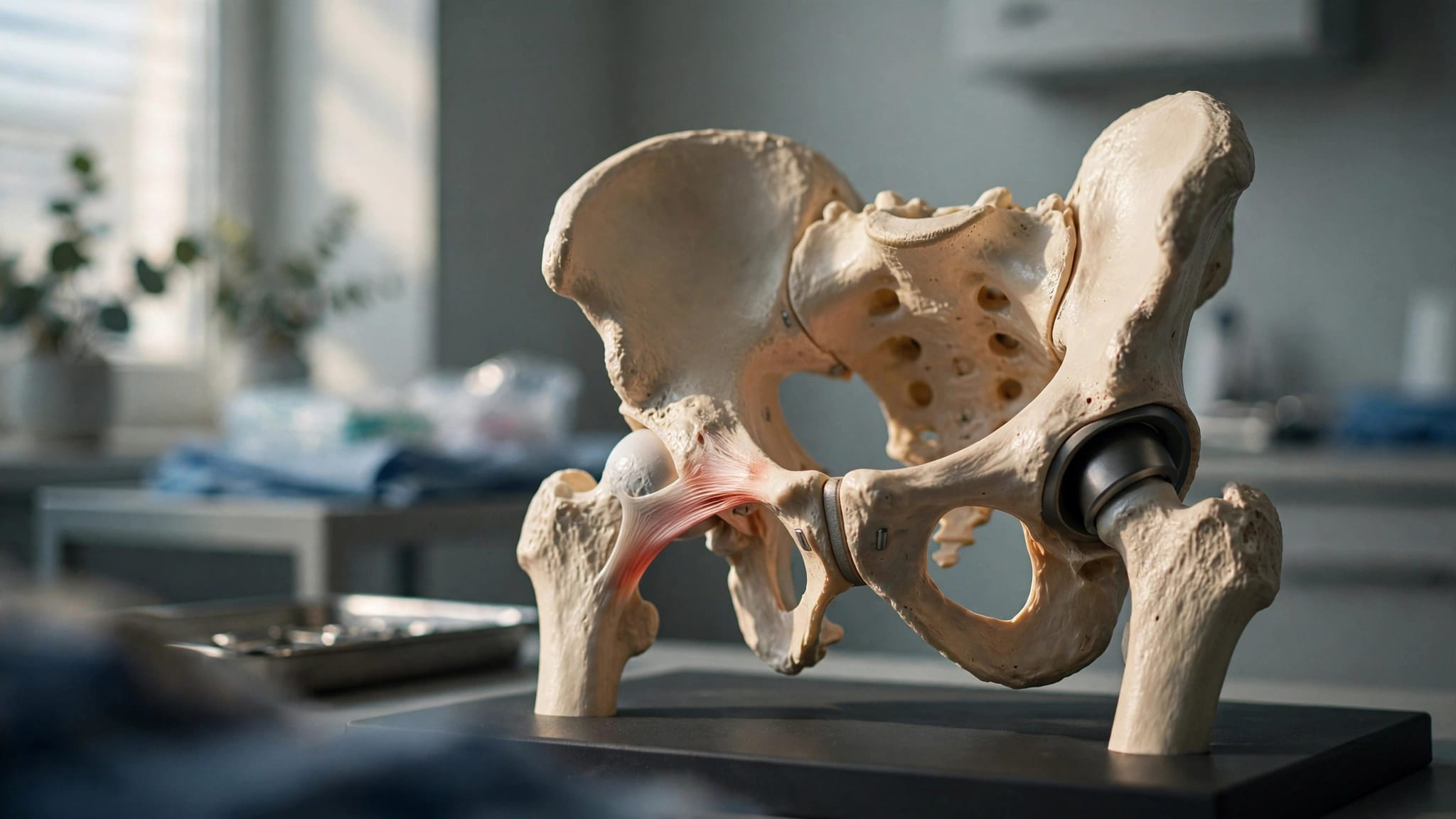
Heterotopic ossification is bone growth in soft tissue around the hip. It can form in muscles, tendons, or connective tissue near the joint after total hip replacement.
The bone does not grow inside the artificial hip components. Instead, it develops around the outside of the joint as part of an abnormal healing response. Small areas may never cause a noticeable problem. Larger areas can interfere with movement.
A 2024 systematic review of 26 studies found that HO appeared on imaging in an average of 28.8% of hip replacements. Yet most of those cases were low grade and did not cause major symptoms.
Most HO is mild
Doctors often describe HO with the Brooker classification, which grades the amount of bone visible on an X-ray. Brooker I shows small islands of bone in the soft tissues. Brooker II shows bone spurs with at least one centimeter between opposite surfaces.
Brooker III means the gap has narrowed to less than one centimeter. Brooker IV describes apparent bony fusion of the hip. Severe grades can limit walking, sitting, dressing, and other daily tasks.
An X-ray finding is not always a problem
A report that mentions heterotopic bone can sound alarming. Still, treatment depends on how you feel and function, not the image alone.
Many people with mild HO have little pain and maintain useful hip motion. In those cases, observation may be more appropriate than an invasive treatment.
Signs That May Point to Heterotopic Bone Growth
HO usually develops during the first several months after surgery. Early on, normal swelling, soreness, and muscle weakness can overlap with its symptoms. The pattern of recovery matters more than one difficult day.
Pain that slowly improves after surgery is expected. However, a hip that becomes progressively stiffer after an early period of improvement should be checked.
Common symptoms of clinically important HO
Symptoms can vary with the amount and location of extra bone. You may notice:
- A growing loss of hip motion, especially when bending, rotating, or stepping into a car.
- Stiffness that feels like a hard block rather than ordinary muscle tightness.
- Pain with walking, transfers, stairs, or putting on socks and shoes.
- A limp that persists because the hip cannot move through a normal stride.
- Difficulty reaching rehabilitation goals despite following the recovery plan.
These signs don't prove that HO is present. Scar tissue, muscle weakness, implant issues, tendon problems, or arthritis in the spine can also affect hip movement.
Symptoms that need prompt medical evaluation
New or worsening pain, stiffness, reduced hip motion, swelling, warmth, or fever should be evaluated promptly. These symptoms are not always heterotopic ossification and may point to infection, a blood clot, a fracture, or another postoperative concern.
Call your surgeon's office promptly if you develop calf pain or one-sided leg swelling. Chest pain or shortness of breath needs emergency care right away. Do not assume these symptoms are part of routine recovery.
How an Orthopedic Surgeon Confirms the Cause
People often use "heterotopic ossification hip replacement" to describe any stiff or painful recovery. An orthopedic examination and imaging help identify the actual cause.
Your surgeon will ask when symptoms began and whether motion improved before getting worse. They will also check your walking pattern, leg strength, hip range of motion, incision, and areas of tenderness.
X-rays show the location and extent of HO
Standard hip and pelvis X-rays are usually the first test. They can show bone forming around the joint and allow the surgeon to assign a Brooker grade.
In some cases, a CT scan gives a clearer view of the bone's location. This can help with surgical planning when HO lies near nerves, blood vessels, or important muscles.
If bone appears to progress several years after hip replacement, the surgeon may look for another cause. Trauma, infection, implant loosening, or other conditions need consideration rather than assuming it is routine postoperative HO.
Some patients have a higher risk
Previous heterotopic ossification is one of the strongest risk factors. A history of hip trauma, earlier hip surgery, ankylosing spondylitis, hip ankylosis, or hypertrophic osteoarthritis can also raise risk.
Men have a higher reported risk in several studies. Surgical complexity and longer operating time may matter as well. Risk factors guide the discussion, but they do not predict one person's outcome with certainty.
Preventing HO in Higher-Risk Patients
Questions about heterotopic ossification hip replacement prevention should come up before surgery when you have a history of HO or another known risk factor. Prevention works best around the time of surgery, before mature bone develops.
The surgical approach can affect HO rates, although it is only one consideration. A recent review found higher reported rates after traditional and modified direct lateral approaches than after posterolateral or direct superior approaches. Your surgeon should choose an approach based on your anatomy, diagnosis, and the control needed for a safe replacement.
Anti-inflammatory medication may reduce risk
Orthopedic surgeons may prescribe an NSAID after surgery for selected patients at higher risk. Indomethacin, naproxen, ibuprofen, and celecoxib are among the medications used in different protocols.
These drugs can reduce the chance of new heterotopic bone formation. They do not remove established bone. The choice and duration depend on your medical history, kidney function, stomach-ulcer risk, heart health, and blood-thinning medication.
Do not start over-the-counter anti-inflammatory medication without your surgeon's approval. Even common medicines can raise bleeding, kidney, or gastrointestinal risks after surgery.
Radiation is reserved for selected situations
External-beam radiation is another preventive option for patients with a substantial risk of severe HO. It is often considered for someone who has had serious HO before or faces a complex hip procedure.
A radiation specialist gives a carefully planned dose shortly before or after surgery. This is not routine for every primary hip replacement. Your surgical team weighs its potential benefit against your health history and the details of the operation.
Treatment Options When HO Is Already Present
Treatment for heterotopic ossification hip replacement symptoms depends on the severity of your limitation, the stage of healing, and the cause of pain. A mild X-ray finding without functional problems often needs monitoring rather than aggressive treatment.
Follow-up visits let your surgeon compare motion, walking, symptoms, and repeat imaging over time. This helps determine whether the condition is stable.
Observation and symptom-guided care
For Brooker I and many Brooker II cases, observation is common. Your surgeon may recommend activity changes, pain control, and periodic checks while the hip continues to heal.
If an anti-inflammatory medication is safe for you, it may help pain from irritated surrounding tissue. However, medication will not dissolve mature heterotopic bone. It should be part of a plan designed for your health needs.
Physical therapy protects function
Physical therapy can help maintain available motion, rebuild hip strength, improve balance, and reduce a compensating limp. It is most useful when the therapist works within the limits of the hip rather than forcing movement through a painful mechanical block.
Aggressive stretching has not been proven to prevent HO. Therapy cannot remove mature extra bone, but it can help you get the most from the motion you have. Your surgeon may adjust the pace or type of exercise if pain and stiffness are increasing.
Recovery plans vary with strength, balance, pain control, and surgical details. This guide to when PT is needed after hip replacement explains why therapy should fit the patient's progress rather than follow a one-size-fits-all schedule.
When Surgical Removal May Be Appropriate
Surgical excision removes heterotopic bone that causes major pain, a hard block to motion, or serious limits on daily life. It is usually considered for severe, mature HO when nonsurgical care cannot restore acceptable function.
The decision is not based on the Brooker grade alone. A person with a higher-grade X-ray finding may still function well, while another person may struggle with less visible bone in a troublesome location.
Timing matters before excision
Surgeons usually wait until the bone has matured and the condition appears stable. Operating too early can raise the chance that it will return. The right timing varies, because healing rates, symptoms, imaging changes, and surgical history differ.
Before recommending removal, the surgeon also confirms that the replacement components are stable. They will look for infection, loosening, nerve problems, or other reasons the hip may hurt or feel stiff.
Excision has real risks and requires planning
Removing HO can improve movement for carefully selected patients. However, the operation can involve bleeding, infection, nerve injury, wound problems, recurrent bone formation, and persistent stiffness.
The surrounding muscles and soft tissues also need protection. In some cases, surgeons use NSAID medication or radiation after excision to lower recurrence risk. A detailed discussion should cover the expected gain in function, recovery demands, and the risks that apply to you.
A Stiff Hip After Replacement Deserves a Clear Answer
Heterotopic ossification can be a minor X-ray finding or a meaningful barrier to motion. The difference comes down to symptoms, function, imaging, healing progress, and your personal risk factors.
Don't dismiss worsening pain or stiffness as something you must tolerate. A clear orthopedic assessment can identify whether HO is involved and whether observation, therapy, preventive medication, or surgery fits your recovery.
ADDITIONAL ARTICLES